Home
/ Brain Anatomy Labeled - Brain Anatomy Labelled Stock Photo Download Image Now Istock, The anterior lobe, the posterior lobe and the flocculonodular lobe.
Brain Anatomy Labeled - Brain Anatomy Labelled Stock Photo Download Image Now Istock, The anterior lobe, the posterior lobe and the flocculonodular lobe.
Brain Anatomy Labeled - Brain Anatomy Labelled Stock Photo Download Image Now Istock, The anterior lobe, the posterior lobe and the flocculonodular lobe.. It is made up of more than 100 billion nerves that communicate in trillions of connections called synapses. Anatomy wise, the cerebellum is located just behind the pons and medulla of the brainstem, near the fourth ventricle. The lobes are functional segments. This article lists a series of labeled imaging anatomy cases by system and modality. This brain part controls thinking.
The anatomy of the brain is complex due its intricate structure and function. Labeled brain diagram first up, have a look at the labeled brain structures on the image below. Try to memorize the name and location of each structure, then proceed to test yourself with the blank brain diagram provided below. See labeled brain anatomy stock video clips of 27 brain diagram with labels hypothalamus vector brain diagram pons cerebrum and cerebellum brain pons brain anatomy amygdala brain labelled amygdala brain human midbrain diagram pons The forebrain, midbrain and hindbrain.
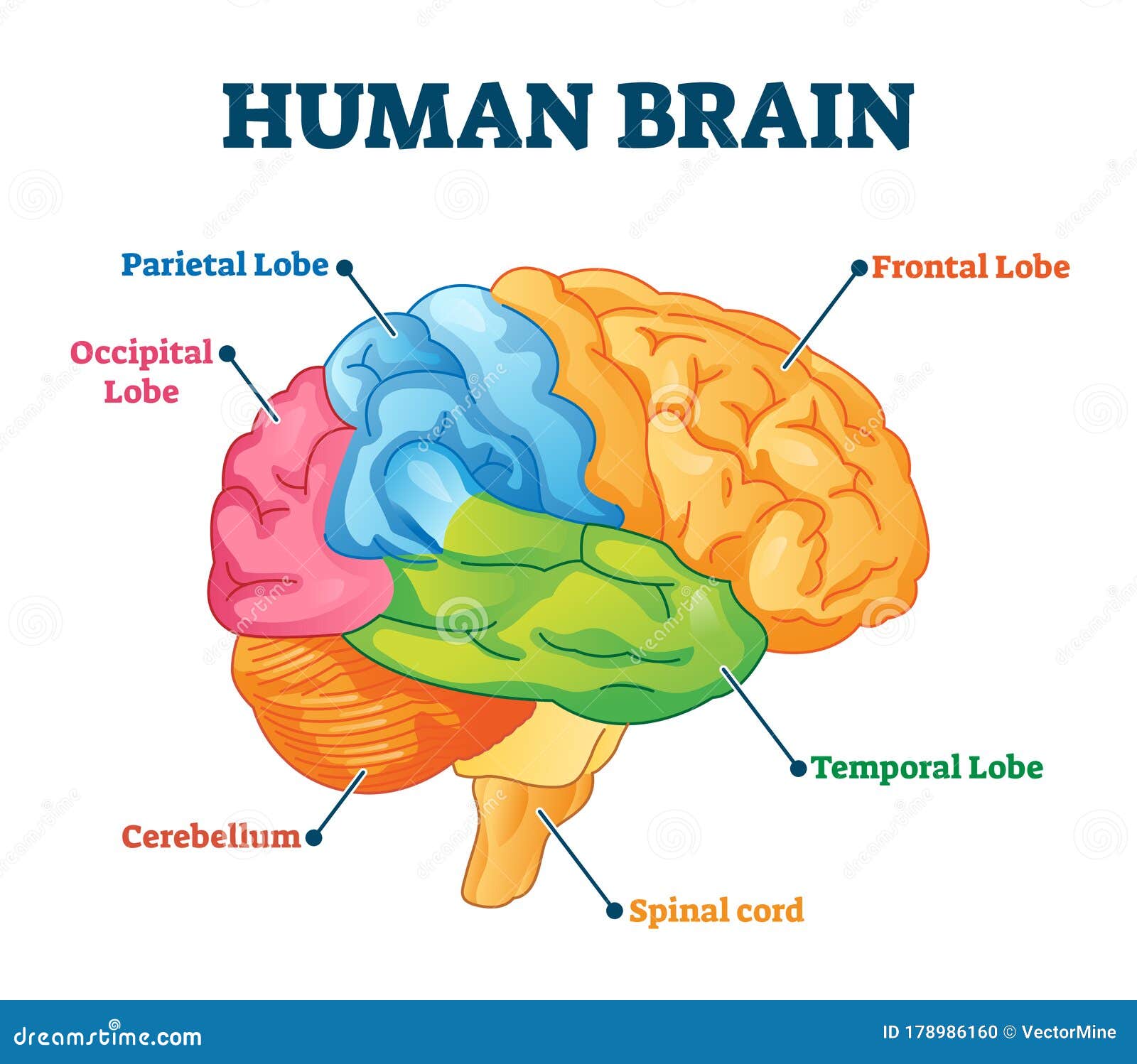
Human Brain Vector Illustration Labeled Anatomical Educational Parts Scheme Stock Vector Illustration Of Explanation Learning 178986160 from thumbs.dreamstime.com It is made up of more than 100 billion nerves that communicate in trillions of connections called synapses. The anterior lobe, the posterior lobe and the flocculonodular lobe. This mri brain cross sectional anatomy tool is absolutely free to use. This brain part controls balance, movement, and coordination. The anatomy of the brain is complex due its intricate structure and function. You are to identify the structures by clicking on the name of the structure. The brain is composed of the complex network of billions of neurons that are arranged in a specific pattern which is vital to the essential functioning of this organ. Anatomy wise, the cerebellum is located just behind the pons and medulla of the brainstem, near the fourth ventricle.
This page presents a comprehensive series of labeled axial, sagittal and coronal images from a normal human brain magnetic resonance imaging exam.
Über 7 millionen englischsprachige bücher. Each brain hemisphere (parts of the cerebrum) has four sections, called lobes: Brainstem responsible for automatic survival reflexes spinal cord controls simple reflexes pathway to neural fibers medulla controls/regulates heartbeat and breathing to and from brain reticular formation helps control arousal, responds to change in monotony. This page presents a comprehensive series of labeled axial, sagittal and coronal images from a normal human brain magnetic resonance imaging exam. Brain anatomy function cheat sheet system or part function misc. Frontal, parietal, temporal and occipital. Try to memorize the name and location of each structure, then proceed to test yourself with the blank brain diagram provided below. The lobes are functional segments. The midbrain helps control eye movement and processes. Use the mouse scroll wheel to move the images up and down alternatively use the tiny arrows (>>) on both side of the image to move the images.>>) on both side of the image to move the images. This article lists a series of labeled imaging anatomy cases by system and modality. See labeled brain anatomy stock video clips of 27 brain diagram with labels hypothalamus vector brain diagram pons cerebrum and cerebellum brain pons brain anatomy amygdala brain labelled amygdala brain human midbrain diagram pons There are different ways of dividing the brain anatomically into regions.
An mri was performed on a healthy subject. Let's use a common method and divide the brain into three main regions based on embryonic development: The lobes are functional segments. Labeled brain anatomy images cortical parcellation of the chimpanzee brain compared to human. Brain anatomy function cheat sheet system or part function misc.
3d Brain from www.brainfacts.org The midbrain, pons and medulla oblongata. Über 7 millionen englischsprachige bücher. See labeled brain anatomy stock video clips of 27 brain diagram with labels hypothalamus vector brain diagram pons cerebrum and cerebellum brain pons brain anatomy amygdala brain labelled amygdala brain human midbrain diagram pons This amazing organ acts as a control center by receiving, interpreting, and directing sensory information throughout the body. We will also learn how cerebrospinal fluid is produced, and how it flows throughout the ventricular system, subarachnoid space, and central canal of the spinal cord. The anterior lobe, the posterior lobe and the flocculonodular lobe. Reviewed by john morrison, patrick hof, and edward lein. This article lists a series of labeled imaging anatomy cases by system and modality.
The anterior lobe, the posterior lobe and the flocculonodular lobe.
We will also learn how cerebrospinal fluid is produced, and how it flows throughout the ventricular system, subarachnoid space, and central canal of the spinal cord. The brain and spinal cord are the two main structures of the central nervous system. An mri was performed on a healthy subject. Labeled brain diagram first up, have a look at the labeled brain structures on the image below. Structure descriptions were written by levi gadye and alexis wnuk and jane roskams. They specialize in various areas of thought and memory, of planning and decision making, and of speech and sense perception. We created a brain atlas that is an interactive tool for studying the conventional anatomy of the normal brain based on a magnetic resonance imaging exam of the axial brain. This tutorial has images in which the structures are labeled. Let's use a common method and divide the brain into three main regions based on embryonic development: Brain anatomy function cheat sheet system or part function misc. It consists of three major parts: The forebrain, midbrain and hindbrain. The anatomy of the brain is often discussed in terms of either the embryonic scheme or the medical scheme.
This amazing organ acts as a control center by receiving, interpreting, and directing sensory information throughout the body. They specialize in various areas of thought and memory, of planning and decision making, and of speech and sense perception. It consists of three major parts: We will use labeled diagrams and lateral views of the brain to learn the anatomy, boundaries, and locations of each ventricle. Dr calum worsley and assoc prof craig hacking et al.
Labeled Dog Brain Anatomy from i.pinimg.com Early on in development, the neural tube forms three outpouchings called the primary brain vesicles. This brain part controls involuntary actions such as breathing, heartbeats, and digestion. The brainstem is the lower extension of the brain, located in front of the cerebellum and connected to the spinal cord. It is made up of more than 100 billion nerves that communicate in trillions of connections called synapses. 11 this brain part controls involuntary actions such as breathing heartbeats and digestion. It serves as a relay station, passing messages back and forth between various parts of the body and the cerebral cortex. This brain part controls thinking. Labeled brain diagram first up, have a look at the labeled brain structures on the image below.
The structure whose name is clicked will be identified in the image by an arrow.
The brainstem is the lower extension of the brain, located in front of the cerebellum and connected to the spinal cord. Use the mouse scroll wheel to move the images up and down alternatively use the tiny arrows (>>) on both side of the image to move the images.>>) on both side of the image to move the images. Download 132 brain anatomy labeled stock illustrations, vectors & clipart for free or amazingly low rates! The anatomy of the brain is often discussed in terms of either the embryonic scheme or the medical scheme. Label the brain anatomy diagram the brain read the definitions below, then label the brain anatomy diagram. Dr calum worsley and assoc prof craig hacking et al. Labeled brain anatomy images cortical parcellation of the chimpanzee brain compared to human. We will use labeled diagrams and lateral images of the brain (side views) to walk through each lobe of the cerebrum. Reviewed by john morrison, patrick hof, and edward lein. See labeled brain anatomy stock video clips of 27 brain diagram with labels hypothalamus vector brain diagram pons cerebrum and cerebellum brain pons brain anatomy amygdala brain labelled amygdala brain human midbrain diagram pons The structure whose name is clicked will be identified in the image by an arrow. The prosencephalon, mesencephalon, and rhombencephalon will form the forebrain, midbrain, and hindbrain, respectively. The brain is one of the largest and most complex organs in the human body.
